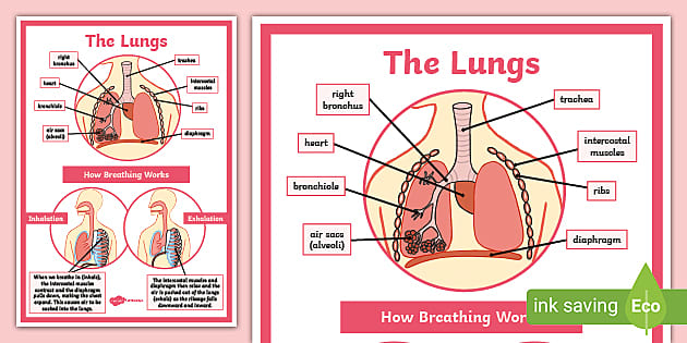
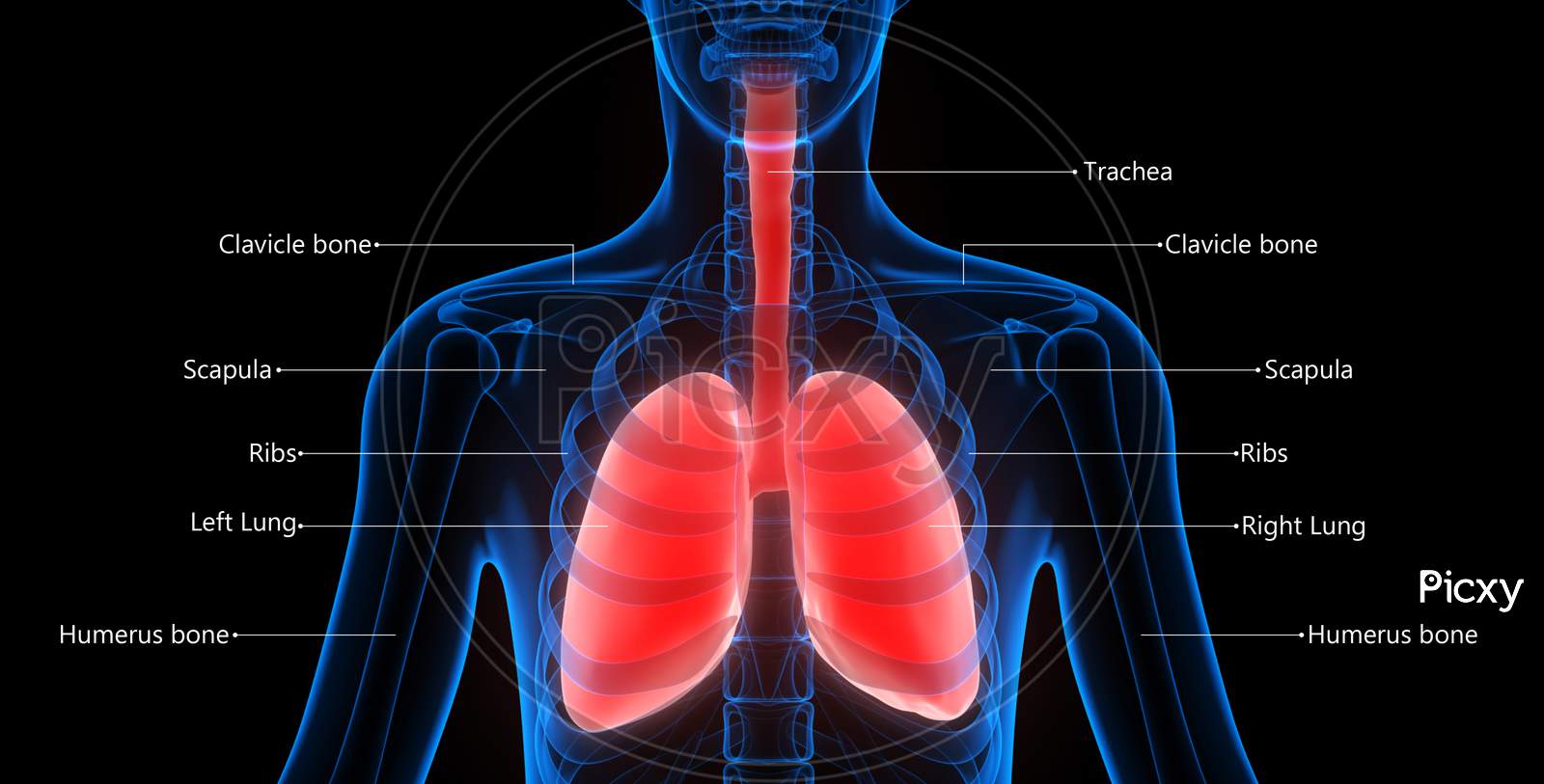
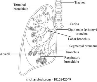
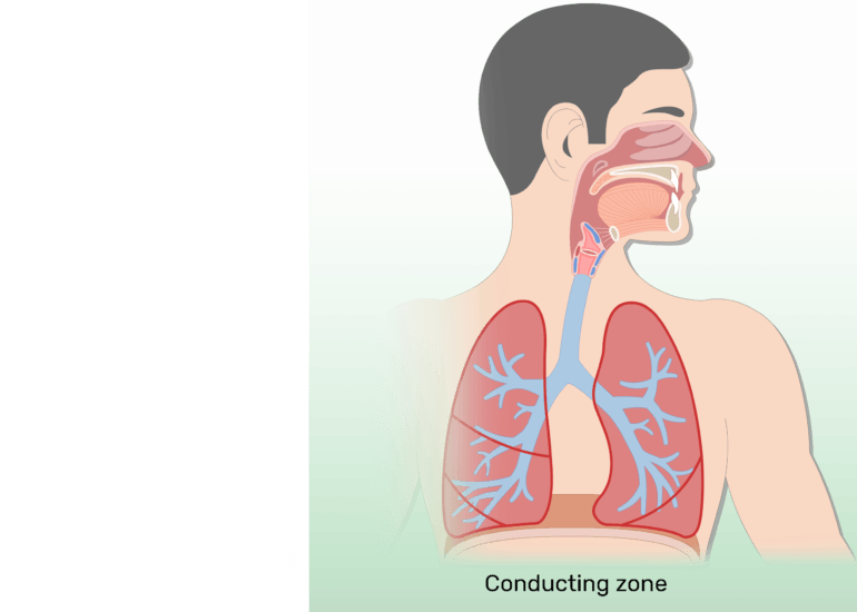
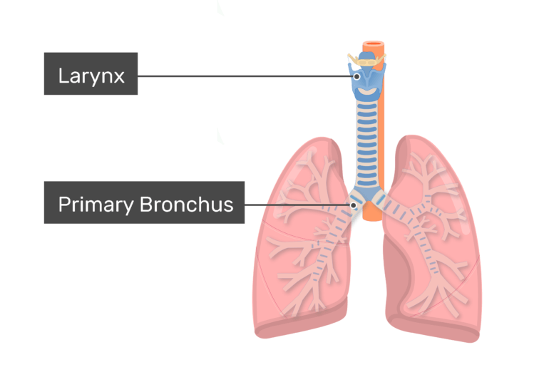
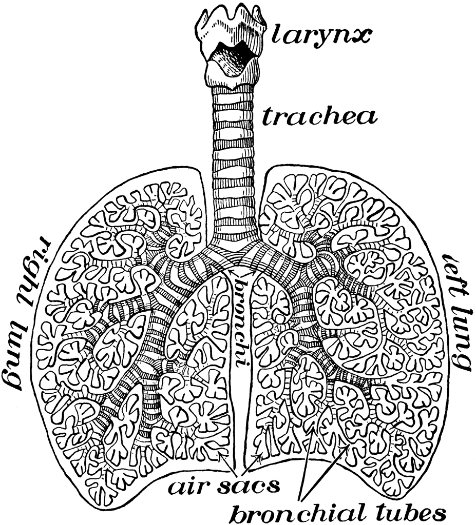
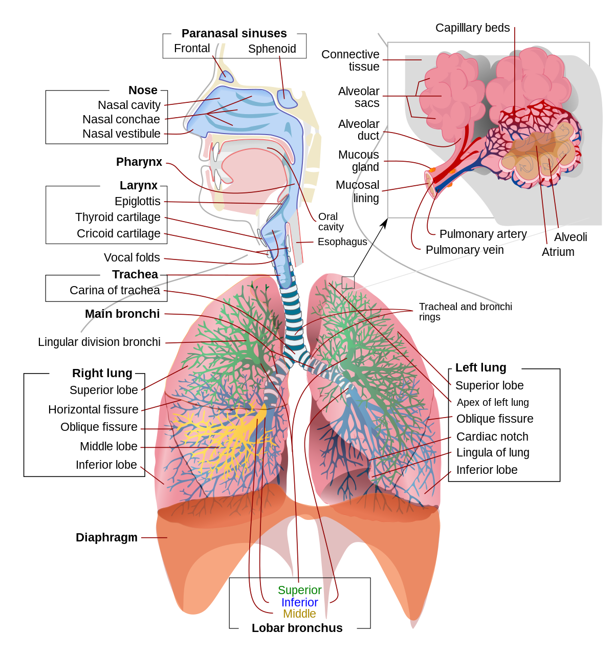
43 diagram of the lungs with labels
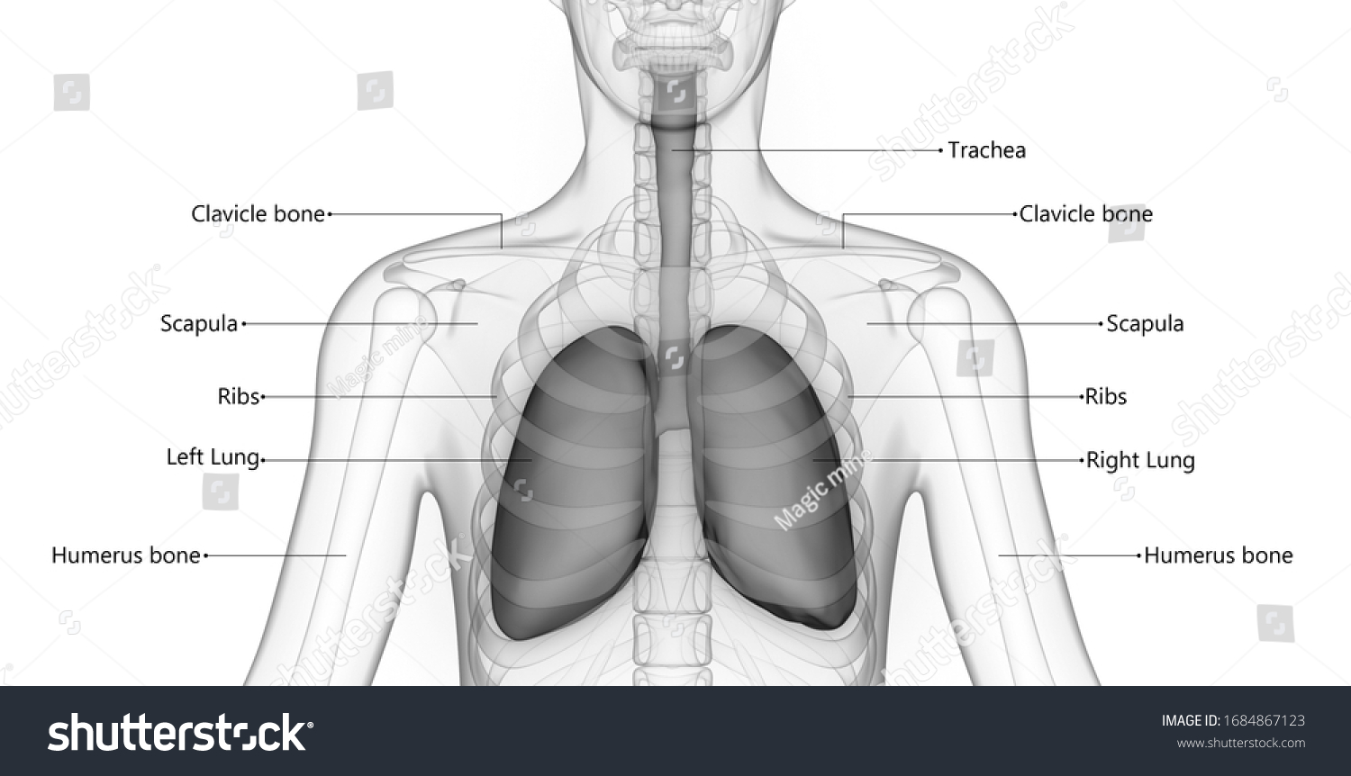
Lungs: Anatomy, Function, and Treatment - Verywell Health Looking at the lungs from the front they lie right above the collarbone and go halfway down the rib cage, although the back of the lungs are slightly longer, ending just above the last rib, while the pleura extends down the entirety of the rib cage. Together with your heart, the lungs take up almost the entire width of the rib cage. 4 Fully Labelled Diagram Alveolus Lungs Showing Stock ... - Shutterstock High Usage score High usage Superstar Shutterstock customers love this asset! Stock Vector ID: 369984683 Fully labelled diagram of the alveolus in the lungs showing gaseous exchange. Vector Formats EPS 1114 × 800 pixels • 3.7 × 2.7 in • DPI 300 • JPG Vector Contributor S Steve Cymro Categories: Science , Education
Diagram Of The Respiratory System With Labels Pictures, Images ... - iStock A medical diagram showing the lobes of the lungs (organ of the respiratory system) with text labels. Human Lungs Diagram Cross section & anterior view of the human lungs. The digestive system The human digestive system medical illustration with internal organs Human Respiratory System Lungs Label Design Anatomy

Diagram of the lungs with labels
Can you label the lungs? Quiz - PurposeGames.com Facial Muscles 10p Image Quiz. Diagram of the Pelvic Girdle 8p Image Quiz. Back Muscles 3p Image Quiz. Labeling the Cochlea 7p Image Quiz. The pancreas 9p Image Quiz. Can you label the eye? 13p Image Quiz. Name the bones of the Skeleton (Posterior) 15p Image Quiz. Labeling the Layers of the Epidermis 12p Image Quiz. Lungs Diagram - Structure, Working, Importance and Facts Lungs are bag-like organs or body parts that allow you to breathe. They are a part of the respiratory system of the human body. Lungs are found in all animals that have a backbone and breathe air. When an animal inhales (breaths in), oxygen-rich air enters the lungs. Carbon dioxide and water vapour are ejected from the lungs when an animal ... The Lungs - Position - Structure - TeachMeAnatomy The lungs are roughly cone shaped, with an apex, base, three surfaces and three borders. The left lung is slightly smaller than the right - this is due to the presence of the heart. Each lung consists of: Apex - The blunt superior end of the lung. It projects upwards, above the level of the 1st rib and into the floor of the neck.
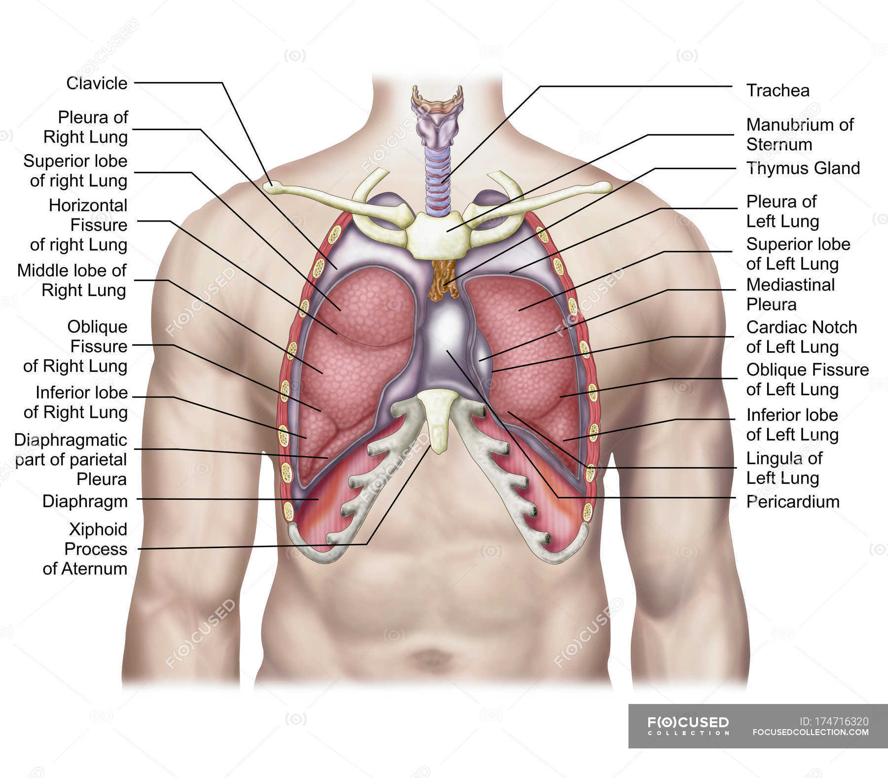
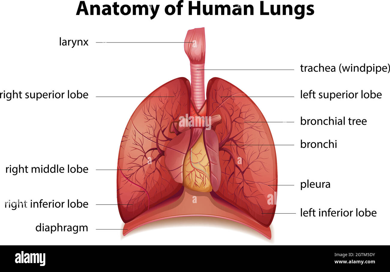
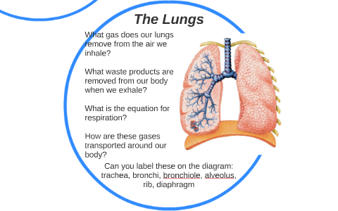
Diagram of the lungs with labels. parts of the lungs labeled anatomy lung labeled pricing netterimages. Circulatory System Diagram - Types Of Circulatory System Diagram . circulatory system diagram circulation arteries cardiovascular systemic veins human types blood diagrams smartdraw body anatomy printable visual teach. Adult Male Diagram Template Clip Art At Clker.com - Vector Clip Art Label the Lungs Diagram | Quizlet ... middle lobe of right lung ... inferior lobe of right lung ... superior lobe of left lung ... left main (primary) bronchus ... lobar (secondary) bronchus ... segmental (tertiary) bronchus ... inferior lobe of left lung ... Sets found in the same folder Lab: Heart Model 34 terms enthusiastic_crafter Bi 233: Labeling the Larynx 21 terms Lungs Diagram - Human Lungs Anatomy - BYJUS The right lung comprises three lobes - inferior, middle and superior lobe that are distinguished by an oblique and deep horizontal fissure. The left lung has two lobes separated by an oblique fissure. The apexes of the lungs expand above the first rib, while both lungs in the thorax rest with their bases on the diaphragm. 9,094 Lung Diagram Images, Stock Photos & Vectors | Shutterstock 9,094 lung diagram stock photos, vectors, and illustrations are available royalty-free. See lung diagram stock video clips Image type Orientation Color People Artists More Sort by Popular Healthcare and Medical Anatomy Diseases, Viruses, and Disorders lung respiratory system medicine organ pulmonary alveolus human body lung cancer of 91
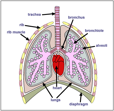
Labeled diagram of the lungs/respiratory system. - SERC View Original Image at Full Size Labeled diagram of the lungs/respiratory system. Image 37789 is a 1125 by 1408 pixel PNG Uploaded: Jan10 14 Last Modified: 2014-01-10 12:15:34 Permanent URL: The file is referred to in 1 page Airborne Microbes Diagram Of The Lungs With Labels Labeling Of The Lungs Label The Lungs ... Diagram Of The Lungs With Labels Labeling Of The Lungs Label The Lungs Diagram Diagram Of Lungs With. By admin Apr 15, 2019. Share this page . Post navigation. Lung Lobectomy: What you need to know . By admin. Related Post. Leave a Reply Cancel reply. You must be logged in to post a comment. Diagram of the Lungs - Etsy Check out our diagram of the lungs selection for the very best in unique or custom, handmade pieces from our prints shops. Lung Anatomy, Function, and Diagrams - Healthline These include the ribs around the lungs and the dome-shaped diaphragm muscle below them. 3-D model of the lungs The lungs are surrounded by your sternum (chest bone) and ribcage on the front...
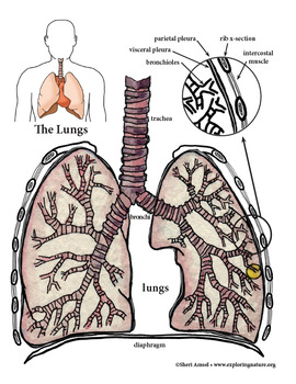
label of the lungs Respiratory system labeled diagrams lungs bio key july. Respiratory system parts epiglottis cavity nasal diaphragm pharynx trachea ppt powerpoint presentation lungs larynx ... Clear lungs & red label chinese herbal 60 capsules. Copd wonderlabs. Unit 4: the respiratory system - douglas college human anatomy & physiology ii (2nd ed.) Anatomy of the Lung | SEER Training - National Cancer Institute Anatomy of the Lung. The lungs are the major organs of the respiratory system, and are divided into sections, or lobes.The right lung has three lobes and is slightly larger than the left lung, which has two lobes.. The lungs are separated by the mediastinum.This area contains the heart, trachea, esophagus, and many lymph nodes. The lungs are covered by a protective membrane known as the pleura ... How to draw and label a lung | step by step tutorial - YouTube How to draw and label a lung | step by step tutorial 76,710 views Premiered Sep 4, 2019 633 Dislike Share Adimu Show 25.8K subscribers A beautiful drawing of Lung. And it will teach you to... Labeled Diagram of the Human Lungs - Bodytomy Given below is a labeled diagram of the human lungs followed by a brief account of the different parts of the lungs and their functions. Each lung is enclosed inside a sac called pleura, which is a double-membrane structure formed by a smooth membrane called serous membrane.
Lobes of the Lung - SmartDraw Venn Diagram Wireframe Lobes of the Lung Create healthcare diagrams like this example called Lobes of the Lung in minutes with SmartDraw. SmartDraw includes 1000s of professional healthcare and anatomy chart templates that you can modify and make your own. 4/22 EXAMPLES EDIT THIS EXAMPLE Text in this Example: Lobes of the Lung
Axial view of labeled lung volumes. Fig. 19: Difference in labels of ... Download scientific diagram | Axial view of labeled lung volumes. Fig. 19: Difference in labels of the ground-truth and the labels generated from the adapted mesh. from publication: Lung lobe ...
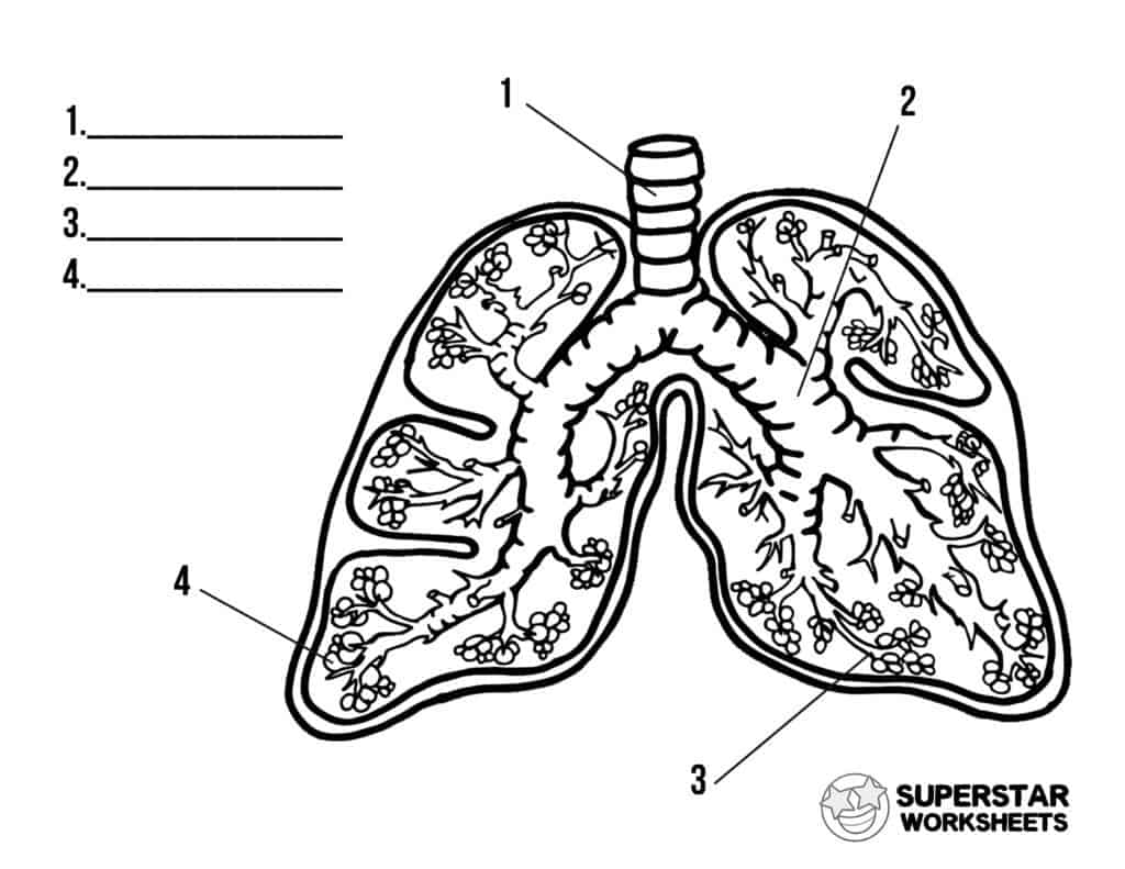
Label Lungs Diagram Printout - Enchanted Learning Label the lungs' lobes, the cardiac notch, and the trachea, larynx, and diaphragm. Instructions For the Student: Read the definitions below, then label the lung anatomy diagram. Extra Information Word Bank bronchial tree: the system of airways within the lungs, which bring air from the trachea to the lung's tiny air sacs (alveoli).
labeled diagram of lungs lung labeled anatomy right lungs labels respiratory system human heart label body blood models physiology medical veins nursing arteries labeling Free Diagrams Of The Lungs | 101 Diagrams lungs diagram unlabeled diagrams pinstake via Ureters: Anatomy, Innervation, Blood Supply, Histology | Kenhub
Lungs (Human Anatomy): Picture, Function, Definition, Conditions - WebMD The lungs are a pair of spongy, air-filled organs located on either side of the chest (thorax). The trachea (windpipe) conducts inhaled air into the lungs through its tubular branches, called...
Lung Diagram Labeled | EdrawMax Template in the following lung labeled diagram, we have shown thyroid cartilage, cricoid cartilage, tracheal cartilage, apex, left upper lobe, hilum, left bronchus, oblique fissure, bronchioles, left lower lobe, base of lung, cardiac notch, right lower lobe, oblique fissure, right middle lobe, horizontal fissure, right bronchus, right upper lobe, and …
Diagram Lungs Illustrations & Vectors - Dreamstime Diagram Lungs Stock Illustrations - 2,750 Diagram Lungs Stock Illustrations, Vectors & Clipart - Dreamstime Diagram Lungs Illustrations & Vectors Most relevant Best selling Latest uploads Within Results People Pricing License Media Properties More Safe Search 2,750 diagram lungs illustrations & vectors are available royalty-free. Next page
Labeled Diagram of the Human Lungs - YouTube Labeled Diagram of the Human Lungs
A Labeled Diagram of the Human Heart You Really Need to See The human heart resembles the shape of an upside-down pear, weighing between 7-15 ounces, and is little larger than the size of the fist. It is enclosed in a bag-like structure called the pericardium, and is located between the lungs, that is in the middle of the chest, behind and slightly to the left of the sternum or breast bone.
labeled diagram of the lungs The Heart Diagrams Labeled And Unlabeled . unlabeled circulatory unlabelled. How The Lungs Work - Amy And Pals amyandpals.com. system respiratory diagram unlabelled human digestive lungs blank lung unlabeled clip clipart clker guion rrt ma lee md cliparts. Copd Cartoons, Illustrations & Vector Stock Images - 1827 Pictures To
Heart Diagram with Labels and Detailed Explanation - BYJUS Well-Labelled Diagram of Heart The heart is made up of four chambers: The upper two chambers of the heart are called auricles. The lower two chambers of the heart are called ventricles. The heart wall is made up of three layers: The outer layer of the heart wall is called epicardium. The middle layer of the heart wall is called myocardium.
The Lungs - Position - Structure - TeachMeAnatomy The lungs are roughly cone shaped, with an apex, base, three surfaces and three borders. The left lung is slightly smaller than the right - this is due to the presence of the heart. Each lung consists of: Apex - The blunt superior end of the lung. It projects upwards, above the level of the 1st rib and into the floor of the neck.
Lungs Diagram - Structure, Working, Importance and Facts Lungs are bag-like organs or body parts that allow you to breathe. They are a part of the respiratory system of the human body. Lungs are found in all animals that have a backbone and breathe air. When an animal inhales (breaths in), oxygen-rich air enters the lungs. Carbon dioxide and water vapour are ejected from the lungs when an animal ...
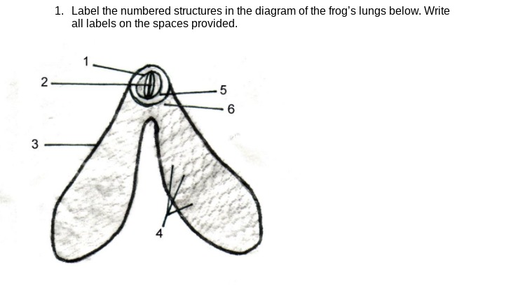
Can you label the lungs? Quiz - PurposeGames.com Facial Muscles 10p Image Quiz. Diagram of the Pelvic Girdle 8p Image Quiz. Back Muscles 3p Image Quiz. Labeling the Cochlea 7p Image Quiz. The pancreas 9p Image Quiz. Can you label the eye? 13p Image Quiz. Name the bones of the Skeleton (Posterior) 15p Image Quiz. Labeling the Layers of the Epidermis 12p Image Quiz.
















Post a Comment for "43 diagram of the lungs with labels"