38 heart structure and labels
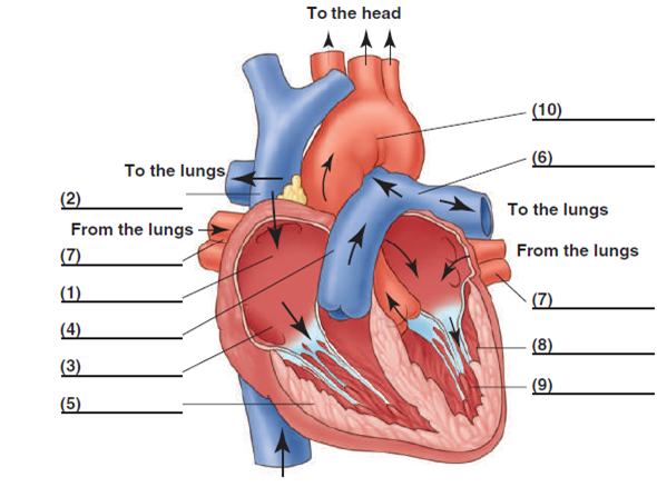
Heart Labeling Quiz: How Much You Know About Heart Labeling? Here is a Heart labeling quiz for you. The human heart is a vital organ for every human. The more healthy your heart is, the longer the chances you have of surviving, so you better take care of it. Take the following quiz to know how much you know about your heart. Questions and Answers. 1. A Diagram of the Heart and Its Functioning Explained in Detail The heart blood flow diagram (flowchart) given below will help you to understand the pathway of blood through the heart.Initial five points denotes impure or deoxygenated blood and the last five points denotes pure or oxygenated blood. 1.Different Parts of the Body ↓ 2.Major Veins ↓ 3.Right Atrium ↓ 4.Right Ventricle ↓ 5.Pulmonary Artery ↓ 6.Lungs
heart diagram and labels heart anatomy lab pig physiology human labeled dissection sheep posterior pigs salvo kctcs bluegrass district edu. Related Items . heart worksheet diagram blank label worksheets sparklebox anatomy teaching simple labelled grade science diagrams classroom coloured related ipc items supplies. Label The Heart Quiz www ...
Heart structure and labels
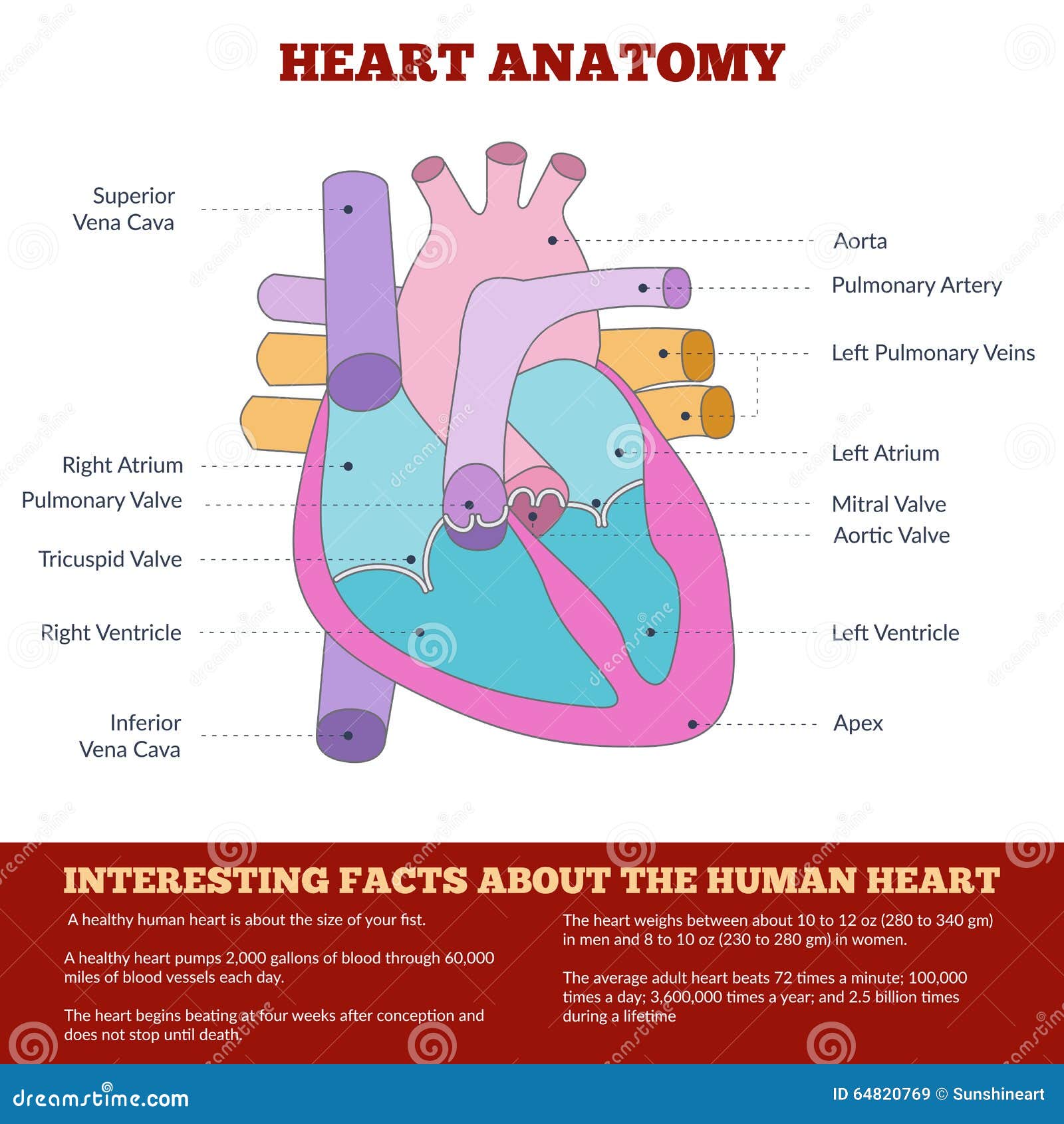
heart | Structure, Function, Diagram, Anatomy, & Facts | Britannica heart, organ that serves as a pump to circulate the blood. It may be a straight tube, as in spiders and annelid worms, or a somewhat more elaborate structure with one or more receiving chambers (atria) and a main pumping chamber (ventricle), as in mollusks. In fishes the heart is a folded tube, with three or four enlarged areas that correspond to the chambers in the mammalian heart. Structure Of The Heart | A-Level Biology Revision Notes The heart is a hollow muscular organ that lies in the middle of the chest cavity. It is enclosed in the pericardium, which protects the heart and facilitates its pumping action. The heart is divided into four chambers: The two atria (auricles): these are the upper two chambers. They have thin walls which receive blood from veins. Human Heart - Diagram and Anatomy of the Heart - Innerbody The heart is a muscular organ about the size of a closed fist that functions as the body's circulatory pump. It takes in deoxygenated blood through the veins and delivers it to the lungs for oxygenation before pumping it into the various arteries (which provide oxygen and nutrients to body tissues by transporting the blood throughout the body).
Heart structure and labels. Human Heart (Anatomy): Diagram, Function, Chambers, Location in Body Chambers of the Heart The heart is a muscular organ about the size of a fist, located just behind and slightly left of the breastbone. The heart pumps blood through the network of arteries and... Structure and Function of the Heart - Medical News The heart is the main organ in the circulatory system, the structure is primarily responsible for delivering blood circulation and transportation of nutrients in all parts of the body. This ... Human Heart - Anatomy, Functions and Facts about Heart The human heart is divided into four chambers, namely two ventricles and two atria. The ventricles are the chambers that pump blood and atrium are the chambers that receive the blood. Among which, the right atrium and ventricle make up the "right portion of the heart", and the left atrium and ventricle make up the "left portion of the heart." 5. Heart: Anatomy and Function - Cleveland Clinic What are the parts of the heart's anatomy? The parts of your heart are like the parts of a house. Your heart has: Walls. Chambers (rooms). Valves (doors). Blood vessels (plumbing). Electrical conduction system (electricity). Heart walls Your heart walls are the muscles that contract (squeeze) and relax to send blood throughout your body.
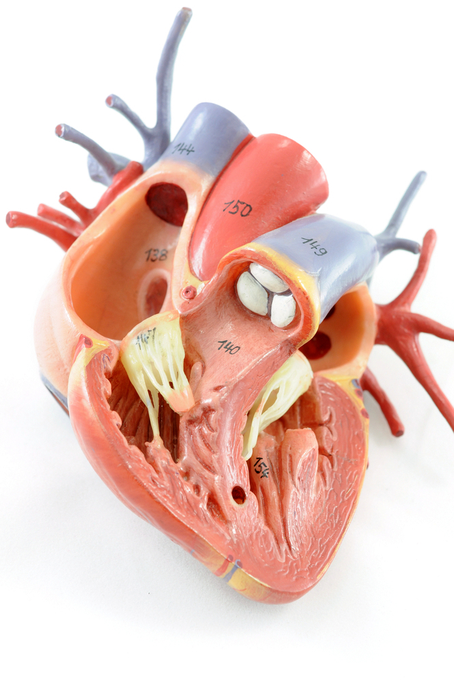
Human Heart Models | Heart Anatomy Models | Vitality Medical The heart model with labels is hand-painted with vivid colors to illustrate the papillary muscles, heart valves, and adjacent structures. Sort By 4 Items Magnetic Heart Model, Life Size, 5 Parts $327.45 View Details Human Heart Model $450.66 - $566.36 View Details Classic Heart Model $81.03 View Details Magnetic Heart Model, Life Size, 5 Part G01 Diagrams, quizzes and worksheets of the heart | Kenhub Worksheet showing unlabelled heart diagrams. Using our unlabeled heart diagrams, you can challenge yourself to identify the individual parts of the heart as indicated by the arrows and fill-in-the-blank spaces. This exercise will help you to identify your weak spots, so you'll know which heart structures you need to spend more time studying ... Label the Heart Diagram | Quizlet Start studying Label the Heart. Learn vocabulary, terms, and more with flashcards, games, and other study tools. Heart anatomy: Structure, valves, coronary vessels | Kenhub Inside, the heart is divided into four heart chambers: two atria (right and left) and two ventricles (right and left).
Heart Diagram with Labels and Detailed Explanation - BYJUS Diagram of Heart. The human heart is the most crucial organ of the human body. It pumps blood from the heart to different parts of the body and back to the heart. The most common heart attack symptoms or warning signs are chest pain, breathlessness, nausea, sweating etc. The diagram of heart is beneficial for Class 10 and 12 and is frequently ... Human Heart Diagram Labeled | Science Trends List Of Heart Structures Heart Chambers Ventricles - The bottom two heart chambers. Atra - The upper two heart chambers. Wall Of The Heart Sinoatrial Node - A collection of tissue that releases electrical impulses and defines the rate of contraction for the heart. Atrioventricular Bundle - The fibers which transmit cardiac impulses. Human Heart - Diagram and Anatomy of the Heart - Innerbody The heart is a muscular organ about the size of a closed fist that functions as the body's circulatory pump. It takes in deoxygenated blood through the veins and delivers it to the lungs for oxygenation before pumping it into the various arteries (which provide oxygen and nutrients to body tissues by transporting the blood throughout the body). Structure Of The Heart | A-Level Biology Revision Notes The heart is a hollow muscular organ that lies in the middle of the chest cavity. It is enclosed in the pericardium, which protects the heart and facilitates its pumping action. The heart is divided into four chambers: The two atria (auricles): these are the upper two chambers. They have thin walls which receive blood from veins.
Label Heart Structure
heart | Structure, Function, Diagram, Anatomy, & Facts | Britannica heart, organ that serves as a pump to circulate the blood. It may be a straight tube, as in spiders and annelid worms, or a somewhat more elaborate structure with one or more receiving chambers (atria) and a main pumping chamber (ventricle), as in mollusks. In fishes the heart is a folded tube, with three or four enlarged areas that correspond to the chambers in the mammalian heart.
Heart of a Frog | ClipArt ETC

The human egg cell explained for egg donors | Altrui
Anatomy Diagram Cartoon Vector | CartoonDealer.com #58232351
Free Heart Diagram Unlabeled, Download Free Heart Diagram Unlabeled png images, Free ClipArts on ...
![Untitled Document [www.stritch.luc.edu]](http://www.stritch.luc.edu/lumen/MedEd/Radio/curriculum/structure/Plain_xray/normal_male_pa_labelled.jpg)
Untitled Document [www.stritch.luc.edu]
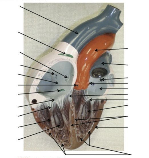
Solved: Label The Following Structures Of The Heart | Chegg.com
Solved: Label the following structures of the heart by writing ... | Chegg.com

34 Label The Heart Diagram - Labels Database 2020

Eclectic Photography Project: Day 144 - strolling around Eureka, CA

Mr. Pite's Room

What are Arteries? - Function & Definition - Video & Lesson Transcript | Study.com
Post a Comment for "38 heart structure and labels"